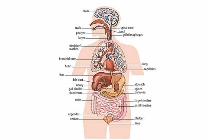
The thyroid gland is one of the most sensitive indicators of microwave influence. Animal experiments show increased activity and/or enlargement of the thyroid at 153 μW / cm2 (Demokidova, 1973) [1], at 100 μW / cm2 (Gabovich et al., 1979; [2] Navakatikian and Tomashevskaya, 1994 [3]), and at 1 μW / cm2 (Dumansky and Shandala, 1973 [4]). Several clinical studies confirm this (Drogichina, 1960 [5]; Sadchikova, 1960 [6]; Smirnova and Sadchikova, 1960 [7]; Baranski and Czerski, 1976 [8]). Smirnova and Sadchikova (1960) [7] states that physiological and even pathological changes in the activity of the thyroid can be detected long before any clinical manifestations of microwave injury. In this study 35 out of 50 persons working with microwave equipment showed abnormal thyroid activity. Drogichina (1960) [5] reports increased thyroid activity in almost all microwave workers examined.
The adrenals are also extremely sensitive to radiation. In animals irradiated for from 2 months up to 2 years, the adrenals are generally enlarged, have an altered ascorbic acid content, increase the secretion of adrenaline and glucocorticoids, and decrease the secretion of testosterone: Chou and Guy at 500 μW / cm2 (Lerner, 1984) [9], Navakatikian and Tomashevskaya (1994) [3] at 100 μW / cm2, Gabovich et al. (1979) [2] at 100 μW / cm2, Dumansky et al. (1982) [10] at 25 μW / cm2, Shutenko et al. (1981) [11] at 10 μW/cm2, Dumansky and Shandala (1973) [4] at 0.06 μW / cm2. With a shorter exposure, Giarola et al. (1971) [12] found a decrease in the mass of the adrenals in chickens at 14-24 μW / cm2. In clinical studies, Sadchikova (1973) [13] noted altered excretion of epinephrine and norepinephrine; Kolesnik et al. noted a decreased blood 17-CHS response to ACTH injection in all 35 workers tested (Baranski and Czerski, 1976 [8]); Hasik, and also Presman, noted increased activity of the adrenal cortex (Dodge, 1969). [14]
Ray and Behari (1990) found a significant decrease in the weight of the spleen, kidney, brain and ovary, and an increase in testicular weight in young rats exposed to 7.5 GHz, 600 μW /cm2, 3 hours a day for 60 days. [15]
Dumansky and Shandala (1973) [4] found increased RNA and DNA in the liver and spleen, and structural changes in the liver, spleen, testes, and brain of white rats and rabbits exposed to 3 cm and 12 cm waves at 0.06 to 101μW / cm2 for 8 to 12 hours a day for 180 days.
Giarola et al. (1971 [12], 1973 [16]) report an enlarged spleen and thymus in baby rats exposed for 35-53 days to 880 MHz, 14 μW / cm2.
Erin’ (1979) [17] reports a 23-83% increase in oxygen tension in renal tissues of adult white rats exposed to 2375 MHz, 50 μW / cm2 for 1-10 days.
Belokrinitsky (1982) [18] observed changes in the biochemistry and ultrastructure of liver, heart, kidney and brain tissue in rats exposed to 12.6 cm waves at intensifies of 5 μW / cm2 and higher for up to 2 months.
50 W / cm2 for 7 hours a day for 10 days caused urine output to fall 15%, and 500 μW / cm2 once for 7 hours had a larger effect (Belokrinitsky and Grin’, 1982 [22]). Elevation of urine pH, protein in the urine, and changes in electrolyte excretion persisted up to 25 days after exposure. Examination of kidney tissue revealed vasodilation, endothelial breakdown, perivascular and pericellular infiltrations, hemorrhage, swelling, partial de-epithelialization along the nephron, and other changes. Histochemical analysis showed decreased cellular glycogen, changes in RNA and DNA concentration, and the appearance of neutral fat droplets. Some of these changes were irreversible, even two months after one 7-hour exposure.
In large clinical studies, Orlova (1960) [19] noted decreased appetite, indigestion, epigastric pain, and enlargement of the liver in irradiated workers, while Gel’fon and Sadchikova (1960) [6] also noted liver enlargement and tenderness in certain patients, with a decreased antitoxic function of the liver in a few. Trinos (1982) [20] noted decreased appetite and indigestion, as well as chronic gastritis, cholecystitis, and decreased gastric acidity, especially in workers exposed to microwaves for more than ten years. Bachurin (1979) [21] also noted chronic gastritis and cholecystitis in workers occupationally exposed to 20-100 μW / cm2.
Bibliograpy
[1] Demokidova, N.K. (1973). On the biological effects of continuous and intermittent microwave radiation. JPRS 63321, p.113.
[2] Gabovich, P.D., Shutenko, O.I., Kozyarin, I.P. and Shvayko, I.I. (1979). Gigiyena i Sanitariya 10:12-14. JPRS 75515, pp. 30-35.
[3] Navakatikian, M.A. and Tomashevskaya, L.A. (1994). Phasic behavioral and endocrine effects of microwaves of nonthermal intensity. In Biological Effects of Electric and Magnetic Fields, D.O. Carpenter and S. Ayrapetyan, eds., Academic Press, N.Y. 1994, pp. 333-342.
[4] Dumansky, J.D. and Shandala, M.G. (1973). The biologic action and hygienic significance of electromagnetic fields of super-high and ultrahigh frequencies in densely populated areas. In Biologic Effects and Health Hazards of Microwave Radiation. Proceedings of an International Symposium, Warsaw, 15-18 Oct. 1973, P. Czerski et al., eds., pp. 289-293.
[5] Drogichina, E.A. (1960). The clinic of chronic UHF influence on the human organism. In The Biological Action of Ultrahigh Frequencies, A.A. Letavet and Z.V. Gordon, eds., Academy of Medical Sciences, Moscow, 1960. JPRS 12471, pp. 22-24.
[6] Sadchikova, M.N. (1960). State of the nervous system under the influence of UHF. In The Biological Action of Ultrahigh Frequencies, A.A. Letavet and Z.V. Gordon, eds., Academy of Medical Sciences, Moscow, 1960, pp. 25-29.
[7] Smirnova, M.I. and Sadchikova, M.N. (1960). Determination of the functional activity of the thyroid gland by means of radioactive iodine in workers with UHF generators. In The Biological Action of Ultrahigh Frequencies, A.A. Letavet and Z.V. Gordon, eds., Academy of Medical Sciences, Moscow, 1960. JPRS 12471, pp. 47-49.
[8] Baranski, S. and Czerski, P. (1976). Biological Effects of Microwaves. Dowden, Hutchinson & Ross, Stroudsburg.
[9] Lerner, E.J. (1984). Biological effects of electromagnetic fields. IEEE Spectrum, May 1984, pp. 57-69.
[10] Dumansky, Y.D., Nikitina, N.G., Tomashevskaya, L.A., Kholyavko, F.R., Zhupakhin, K.S. and Yurmanov, V.A. (1982). Meteorological radar as source of SHF electromagnetic field energy and problems of environmental hygiene. Gigiyena i Sanitariya 2:7-11, 1982a. JPRS 84221, pp. 58-63.
[11] Shutenko, O.I., Kozyarin, I.P. and Shvayko, I.I. (1981). Effects of superhigh frequency electromagnetic fields on animals of different ages. Gigiyena i Sanitariya 10:35-38, 1981. JPRS 84221, pp. 85-90.
[12] Giarola, A.J., Krueger, W.F. and Woodall, H.W. (1971) The effect of a continuous UHF signal on animal growth. 1971 IEEE Inter-national Electromagnetic Compatibility Symposium Record, Phila., July 13-15, 1971, pp. 150-153.
[13] Sadchikova, M.N. (1973). Clinical manifestations of reactions to microwave irradiation in various occupational groups. In Biologic Effects and Health Hazards of Microwave Radiation: Proceedings of an International Symposium, Warsaw, 15-18 Oct., 1973, P. Czerski et al., eds., pp. 261-267.
[14] Dodge, C.H. (1969). Clinical and hygienic aspects of exposure to electromagnetic fields. In Symposium Proceedings. Biological Effects and Health Implications of Microwave Radiation, S. Cleary, ed., Richmond, Va., Sept. 1969, pp. 140-149.
[16] Giarola, A.J., Krueger, W.F., and Neff, R.D. (1973). The growth of animals under the influence of electric and magnetic fields. Health Physics in the Healing Arts, Seventh Midyear Topical Symposium, Health Physics Society, San Juan, P.R., Dec. 11-14, 1972, published March 1973, pp. 502-509.
[17] Erin’, A.N. (1979). Changes in oxygen tension, temperature and blood flow velocity in animal renal tissues during irradiation with low intensity electromagnetic waves in the ultrahigh frequency range. Vrachebnoye Delo 11:110-111, 1979. JPRS 75515.
[18] Belokrinitsky, V.S. (1982). Destructive and reparative processes in hippocampus with long-term exposure to nonionizing microwave radiation. Bulletin of Experimental Biology and Medicine 93(3):89-92, 1982. JPRS 81865, pp. 15-20.
[19] Orlova, A.A. (1960). The clinic of changes of the internal organs under the influence of UHF. In The Biological Action of Ultrahigh Frequencies, A.A. Letavet and Z.V. Gordon, eds., Academy of Medical Sciences, Moscow, 1960. JPRS 12471, pp. 30-35.
[20] Trinos, M.S. (1982). Frequency of diseases of digestive organs in people working under conditions of combined effect of lead and SHF-range electromagnetic energy. Gigiyena i Sanitariya 9:93-94, 1982. JPRS 84221, pp. 23-26.
[21] Bachurin, I. V. (1979) Influence of small doses of electromagnetic waves on some human organs and systems. Vrachebnoye Delo 7:95-97, 1979. JPRS 75515, pp. 36-39.
[22] Belokrinitsky, V.S. and Grin’, A.N. (1982). Nature of morpho-functional renal changes in response to SHF fieldhypoxia combination. Vrachebnoye Delo 1:112-115, 1982. JPRS 84221, pp. 27-31.


